


MIT's Augmented Reality Ultrasound System Helps Novices Match Expert Accuracy
First-time ultrasound users equipped with an augmented reality headset performed nearly as well as trained sonographers and physicians on spatial tasks, according to research published today in Nature Communications Engineering. The MIT team's augmented reality ultrasound system works not by improving the underlying scan, but by eliminating the step that makes ultrasound hard to read in the first place, MIT News reported today.
The study involved 18 participants and tissue phantoms in a controlled lab setting. It is not a clinical trial. But the mechanism it demonstrates is specific: remove the mental 2D-to-3D reconstruction burden, and the performance gap between novices and experts narrows sharply.
How MIT's 3D augmented reality ultrasound system works
Standard ultrasound hands the operator a flat 2D image and asks them to mentally assemble multiple cross-sectional slices into a coherent picture of tissue in real time. That reconstruction task is what makes ultrasound interpretation difficult to learn, per MIT News.
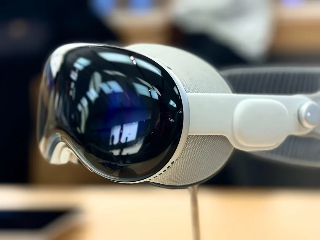
The MIT system, called AR-VIU (augmented real-time volumetric imaging in ultrasound), handles that reconstruction automatically. A probe slightly smaller than a deck of cards transmits raw data via a chirped acquisition system. That data is compressed and streamed into Unreal Engine, which converts the voxel information into a live 3D volumetric model with no loss of information, visible through a VR headset, per MIT News. Where a standard operator reads a flat slice on a monitor, an AR-VIU user sees the scanned structure as a three-dimensional shape in space.
Because AR-VIU uses fewer transducer elements than a conventional 3D ultrasound system, it requires less power and costs less to build, MIT News notes. That matters for anything that needs to move out of a research lab.
Lead author Shrihari Viswanath described the change as providing "3D visual context" that makes ultrasound "significantly easier for novices to understand." Senior author Canan Dagdeviren put it more plainly: "less brain drain." The MIT report frames the gain as one of visualization rather than image capture. The scan itself does not change. What changes is what the operator has to do with it.
What the study found, and where it stops
Nine ultrasound professionals (practicing sonographers and physicians) and nine people with no prior ultrasound experience each performed tasks under two conditions: standard 2D imaging on a monitor, then AR-VIU through a headset. In one set of experiments, participants identified objects embedded in gelatin inside an opaque container (a spring, a ball, a screw). In a second set, they marked the location of a tissue-mimicking gel phantom, per MIT News.
AR-VIU significantly improved object identification and localization accuracy across all 18 participants. Under standard 2D imaging, experts performed much better than novices. Under AR-VIU, novices performed nearly as well as experts on these lab tasks, MIT News reports. After the experiments, most novice participants said they preferred AR-VIU and found the tasks easier to complete.
That result is striking but bounded. The study measures spatial identification and localization accuracy in a lab with tissue phantoms. It does not measure diagnostic accuracy, whether a clinician could correctly characterize tissue pathology, or whether novice gains hold in a real patient exam where anatomical variability, patient motion, and time pressure all apply. The MIT team acknowledges they are still working to improve imaging resolution and conduct further accuracy testing before the system could be considered for clinical use, per MIT News.
Easier to read spatially is not the same as easier to interpret diagnostically. That gap is where the next phase of testing lives.
Where the finding has the most immediate use
The MIT team identifies two near-term applications: accelerating training for ultrasound technicians and other providers, and guiding needle placement for biopsies, where the margin for spatial error is directly consequential, per MIT News. Dagdeviren described the clinical upside as procedures that would be "less time-consuming, more accurate," giving providers confidence "they wouldn't have to wonder if they missed anything."
Training is probably the more immediate application. A PubMed-indexed review of AI assistance in pediatric point-of-care ultrasound, published earlier this year, found a consistent pattern: technology that reduces interpretation burden delivers its largest performance gains for less experienced users and shows the greatest promise in settings where specialist access is limited, according to the review. That work focuses specifically on AI-assisted pediatric POCUS, a different technology than AR-based volumetric imaging, but the common thread is relevant: support tools help novices most, and that advantage matters most where expert backup is scarce.
For procedural guidance, a separate mixed-reality biopsy navigation study published two months ago took a different approach entirely, focusing on displaying real-time ultrasound at its true anatomical position in a head-mounted display, co-registered with pre-procedural CT or MRI. That study's markerless tracking method held target registration error within 3.41 mm at 95% probability and showed strong potential to reduce needle insertion attempts compared to conventional ultrasound guidance alone, per PubMed. The value proposition is different from MIT's work, precision over training accessibility, but both point to the same structural limitation: standard ultrasound creates spatial ambiguity at exactly the moment accuracy matters most.
What comes next
The clearest finding from the MIT study is not that experts improved with AR-VIU. It is that novices, using the system, performed nearly as well as experts on spatial tasks where 2D imaging produced a wide performance gap, per MIT News. The mechanism is specific and the lab result is reproducible. Whether it holds under clinical conditions is a different question.
That is what real-patient testing needs to answer: whether the novice gains survive when anatomy varies, when patients move, when there is no gelatin phantom and no controlled task, and when the outcome is a diagnostic call rather than a localization score. The broader pattern from AI-assisted ultrasound research suggests the gains will be largest for less experienced users in lower-resource settings, per the POCUS review. But the pattern is not a guarantee.
If clinical validation holds, the question stops being how to train more ultrasound experts and starts being how to build interfaces that make the core skill more portable. That is a more tractable problem. The prototype makes a reasonable case for where to start.


Comments
Be the first, drop a comment!